
Fluorescence & Imaging
We personally find you the right new, used and refurbished fluorescence & imaging instruments & equipment from verified suppliers.
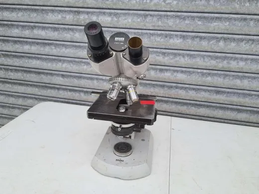
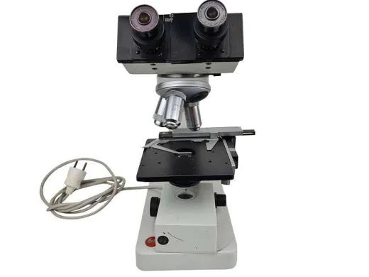
Fluorescence and imaging work turns a labelled sample into a measurable signal, and the equipment choice binds on two things at once: what excites the fluorophore and what records the light that comes back. A confocal has to resolve a plane within a thick specimen; a gel imager has to capture a faint band against a dark field; a western blot reader has to separate two overlapping emission channels. Get the pairing of light source, optics and detector wrong and the signal is there but unreadable. The Questpair catalogue carries 16994 listings across imaging systems, microscopes, molecular imagers and the cameras and accessories that complete them, from 163 verified suppliers and distributors.
Updated:
Fluorescence & Imaging product list


Not the exact instrument you need?
Tell our scientists what you are after. We track it down, including off-market supply.
Not the exact instrument you need?
Tell our scientists what you are after. We track it down, including off-market supply.
Showing 1–40 of 7,274 listings
Show 40 more listingsBuying guide
This page spans more categories than any single one of them, so the price range is the widest on the site. Across the work, listings run from EUR 4 to EUR 244.724. The bottom end is accessories and consumables: a filter cube, a slide holder, a gel dryer part, the small pieces that complete a setup already in place. The top end is the research confocal, a scanning head, laser lines, motorised stage and sensitive detector together, which is where the four and five-figure figures sit. Molecular imagers and complete fluorescence microscopes fall between, closer to the top when they arrive as full systems with camera and filters.
Condition is where this catalogue leans heavily one way. Of the listings, 95 percent are used. For optics that is less of a concern than it sounds: a well-kept objective or a confocal scanner does not degrade the way a moving instrument might, and the saving against new is substantial. What matters more is completeness. A microscope is only as capable as its filter cubes, and an imager only reads the dyes its installed filters allow, so check what a listing actually includes before comparing prices.
Where you have to weigh a used system against a new one, the clear difference is price: used and refurbished units list below new. Warranty is often possible and is arranged in consultation for the specific listing, so that is the place to settle it rather than assuming it from the condition alone. Parts and accessories can be sourced via Questpair, which is what makes buying an incomplete system and completing it a workable route.
Types of Fluorescence & Imaging
Fluorescence and imaging is not a single workflow but a set of related ones, held together by one idea: light in, light out, recorded. The equipment this draws on spans microscopes and confocal systems for looking at cells, molecular imagers and gel documentation systems for reading blots and gels, and the cameras, filters and accessories that make either possible. The sections below group the work by what it is trying to see, because that is what decides the kit.
How do microscopy and confocal systems differ for fluorescence?
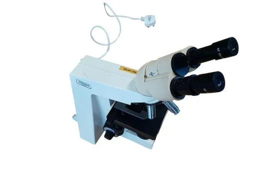
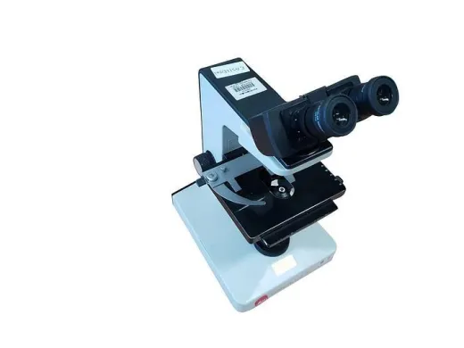
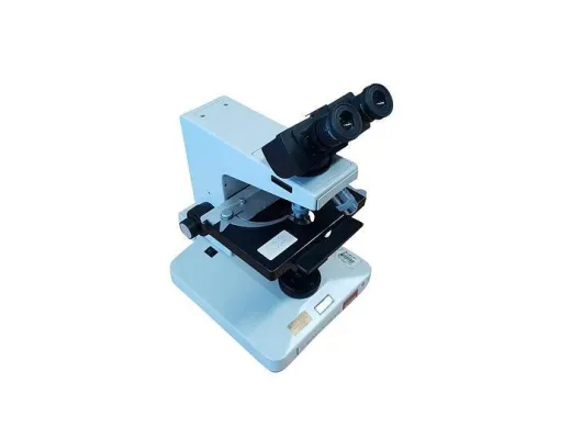
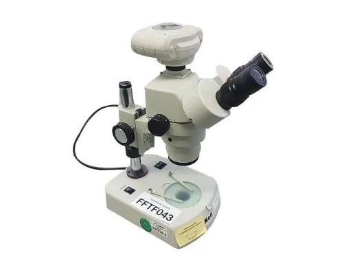
A widefield fluorescence microscope floods the whole field with excitation light and reads the emission on a camera. It is fast and simple, and for thin samples, a monolayer of cells, a smear, a section a few microns deep, it is enough. The limit shows up with thickness: out-of-focus light from above and below the plane washes into the image and the contrast collapses.
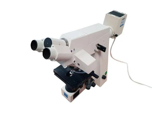
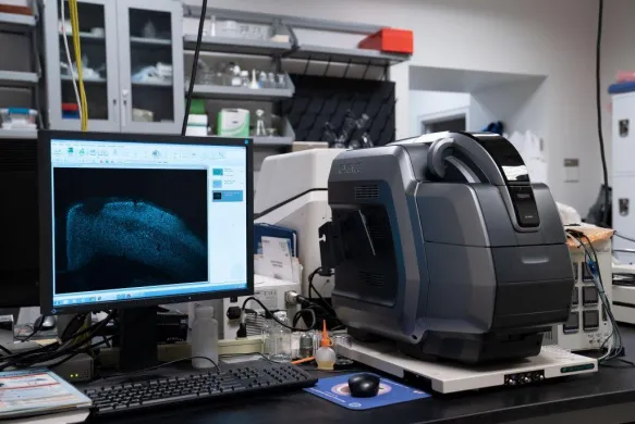
A confocal answers that by rejecting the out-of-focus light. A pinhole in front of the detector admits only light from the focal plane, so the system can build a clean optical section and, by stepping through the sample, a stack. That is what thick specimens and three-dimensional reconstruction need. The cost is complexity: a scanning head, precise focus control and, usually, laser excitation rather than a lamp. On the marketplace both sit under the microscopy listings, and the accessories, objectives, stages, filter sets, often transfer between them.
The practical decision is sample depth against budget. A lab imaging fixed slides rarely needs the confocal; a lab imaging organoids or whole-mount tissue cannot do without one.
Reading blots and gels: what does molecular imaging need?
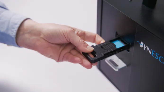
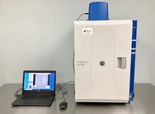
Western blot and gel imaging is a separate branch of the same physics. Here the subject is a membrane or a gel carrying bands, and the signal is often faint. The equipment, molecular imagers and gel documentation systems, is built around a sensitive camera, a dark enclosure to keep stray light out, and a choice of excitation. Fluorescent westerns need the right excitation wavelength and emission filter for each dye; chemiluminescent blots need no excitation at all, just a camera patient enough to integrate a weak glow over a long exposure.
Where two steps constrain each other is the dye-to-filter match. A two-colour western only separates cleanly if the imager carries filters that isolate each emission band, and a system bought for one dye set may not read another. This is the kind of dependency a category page for cameras or imagers cannot state, because it only appears once the detector and the chemistry are considered together.
Gel dryers belong to the same bench: once a gel has been imaged, drying preserves it for storage or autoradiography. They are listed here because they sit at the end of this particular sequence, not because they image anything themselves.
What do cameras and accessories add?
The camera is where much of the sensitivity actually lives. A cooled scientific camera on a fluorescence microscope pulls detail out of a dim sample that a standard sensor would lose in its own noise. On the molecular imaging side, the same choice decides whether a faint band registers at all. Among the listings that specify a type, cameras appear both as part of complete systems and on their own, for fitting to an existing stand.
The accessories, objectives, filter cubes, stages, illuminators, are what let one instrument serve more than one assay. A filter set defines which fluorophores a given microscope can read, so adding a cube often matters more than changing the microscope. Buying these second-hand is where the breadth of this catalogue tells.
Frequently asked questions
What equipment do you need at minimum to start fluorescence imaging?
A light source matched to your fluorophore's excitation, the optics to deliver and collect that light, and a detector to record the emission. For a basic widefield setup that means a fluorescence microscope with the right filter cube and a camera sensitive enough for your sample; for blots it means a molecular imager with a dark enclosure, the correct excitation and emission filters, and a camera.
Which part of a fluorescence setup is the most expensive to set up?
The research confocal, because it combines a scanning head, laser lines, a motorised stage and a sensitive detector in one instrument, which is where the four and five-figure listings sit. A widefield microscope or a molecular imager costs less, and standalone accessories such as filter cubes and stages are at the bottom of the range.
What can be bought used and what is better bought new for imaging work?
Optics tolerate used condition well: a well-kept objective, filter cube or confocal scanner does not wear the way a moving mechanism does, and used and refurbished units list below new. Of the listings, 95 percent are used, so the practical question is less condition than completeness, whether a given system arrives with the filters and camera it needs.
What has to match between the steps in fluorescence imaging?
The fluorophore, the excitation source and the emission filter all have to agree, and this is the dependency that catches buyers out. A two-colour western only separates cleanly if the imager carries filters that isolate each dye's emission band, and a microscope can only read the fluorophores its installed filter cubes allow, so a system bought for one dye set may not read another.
Roughly what does a complete fluorescence and imaging setup cost?
Across this work listings run from EUR 4 to EUR 244.724, the widest range on the site because it spans small accessories through to full research systems. Complete fluorescence microscopes and molecular imagers with camera and filters fall toward the upper part of that range, while a research confocal with laser lines and a motorised stage sits at the top.
Where does cutting the budget cost you later in imaging work?
Skimping on the camera, since much of the sensitivity lives there: a standard sensor loses a dim fluorescence signal or a faint blot band in its own noise, where a cooled scientific camera recovers it. Buying an imager with too narrow a filter set is the other common trap, because it limits which dyes you can ever read without adding filters.
Which imaging equipment is shared with other work on the bench?
Accessories transfer the most: objectives, stages, filter cubes and illuminators often move between a widefield microscope and a confocal, since both sit under the microscopy listings. Cameras appear both inside complete systems and on their own for fitting to an existing stand, and a gel dryer serves any workflow that ends in preserving a gel rather than imaging one.
What should you check on used imaging equipment before buying?
Check what the listing actually includes rather than the headline instrument, because a microscope is only as capable as its filter cubes and an imager only reads the dyes its filters allow. Confirm the excitation sources and emission filters cover your dyes, and where a system is incomplete note that parts and accessories can be sourced via Questpair to finish it.
About Questpair
Questpair is the European marketplace for lab equipment, where AI-prepared cases and personal specialists guide every purchase. We work with 247 verified suppliers across 38 countries to find buyers the right new, used or refurbished instruments, with response within 1 hour and significant savings vs buying new.
Personal · Fast · Reliable · Affordable