
Microscopy & Imaging
We personally find you the right new, used and refurbished microscopy & imaging instruments & equipment from verified suppliers.
Resolving a structure means holding three things steady at once: the optics that form the image, the stage that keeps the specimen still, and the detector that records it before anything drifts. A soft stand and a good objective photograph nothing worth keeping, so the binding constraint is rarely the headline magnification. It is sample preparation, mechanical stability and the match between objective and detector. Questpair lists 18329 items for this work, from benchtop stands through confocal and electron platforms. Specimen handling runs alongside the optics: a sample has to be fixed, sectioned and mounted before it ever reaches the lens, and the quality of that step sets the ceiling on everything after it.
Updated:
Microscopy & Imaging product list


Not the exact instrument you need?
Tell our scientists what you are after. We track it down, including off-market supply.
Not the exact instrument you need?
Tell our scientists what you are after. We track it down, including off-market supply.
Showing 1–40 of 7,467 listings
Show 40 more listingsBuying guide
Prices here run from EUR 4 to EUR 244.724. That spread is wider than on any single category page, because this work covers everything from a box of coverslips to a complete electron platform. At the low end sit the consumables and the small accessories, the slides, the holders, the objectives bought singly. The expensive end belongs almost entirely to the image-forming instruments: confocal systems, and above them the electron and atomic force platforms, where the optics, the vacuum column and the detector are one integrated purchase rather than a stand with parts bolted on. The preparation equipment, processors, microtomes, stainers, tends to fall in the middle, substantial but not in the class of a beam instrument.
Condition matters more at this end of the market than at most. 95 percent of the listings are used. For a stand or a preparation instrument that is often the sensible buy, since the mechanics are mature and a used unit is listed below a new one. For an electron or confocal platform the calculation is tighter, because alignment and detector state carry a cost that a price tag alone does not show, and warranty on any specific unit is arranged in consultation for that listing rather than assumed. Where the decision is close, the thing to read is the individual listing and what it says about condition, not a generic rule. Parts for these instruments can be sourced via Questpair, which widens what is worth considering second-hand.
Types of Microscopy & Imaging
Microscopy and imaging is less a single procedure than a chain that starts at the specimen and ends at a recorded frame. The equipment splits accordingly: preparation tools that turn tissue into a section thin enough to see through, the instruments that form and magnify the image, and the detectors and stages that capture it. What follows walks that chain in the order a sample actually travels it, and names what each step draws on.
From specimen to slide: what does sample prep demand?
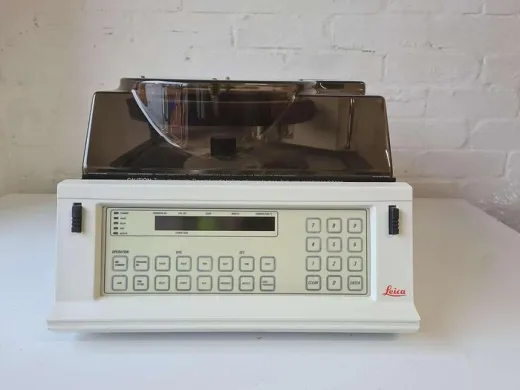
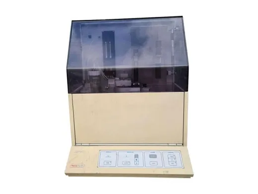
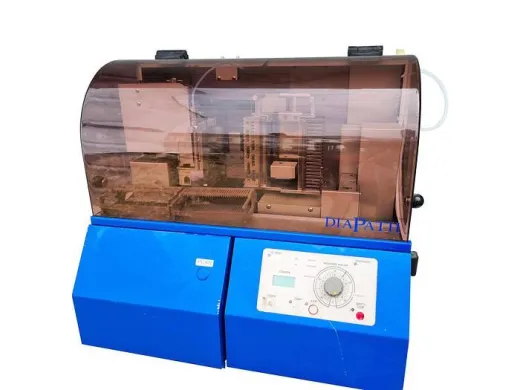
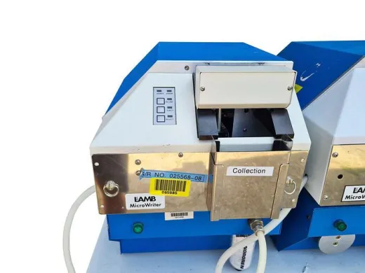
Nothing reaches an objective unprepared. Tissue has to be fixed, dehydrated and infiltrated with a supporting medium before it holds together under a blade, which is the job of a tissue processor and, afterward, an embedding station that sets the block. A microtome then cuts sections measured in single-digit microns, and the ribbon is floated onto coverslips or slides. For anything that will be read by eye or by brightfield, a slide stainer applies the contrast that raw tissue lacks. Two steps constrain each other here in a way no single category page can show: section thickness set at the microtome decides how much stain penetrates, and a block embedded unevenly tears before it ever sections cleanly. The listings carry processors, embedding stations, microtomes and stainers as separate equipment, but in practice they are bought as a sequence, because a gap anywhere in it shows up as artefact at the lens.
Forming the image: which instrument for which structure?
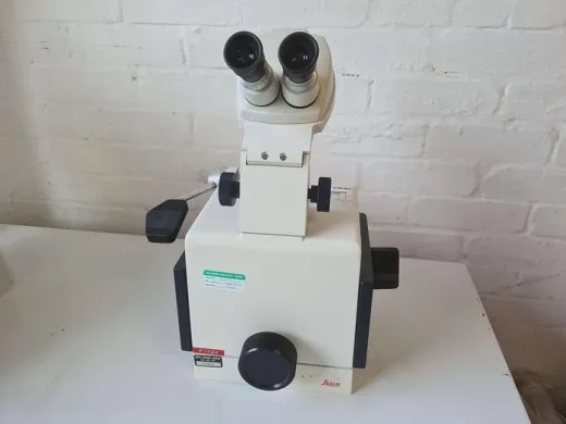
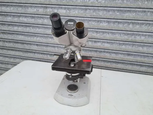
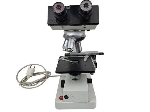
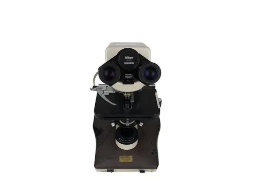
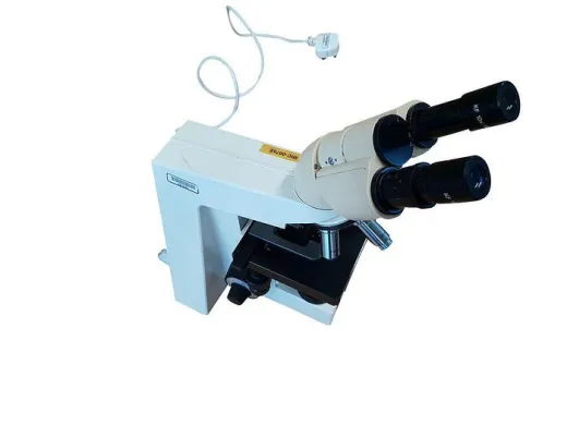
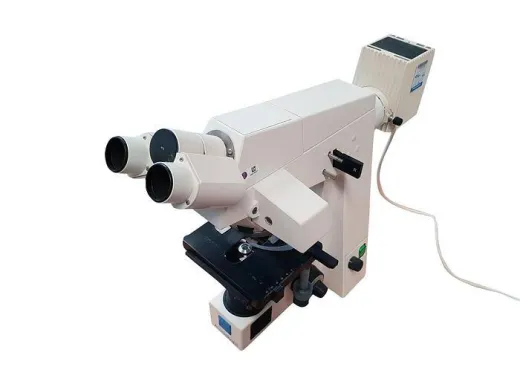
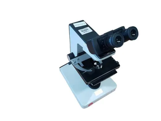
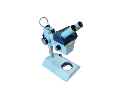
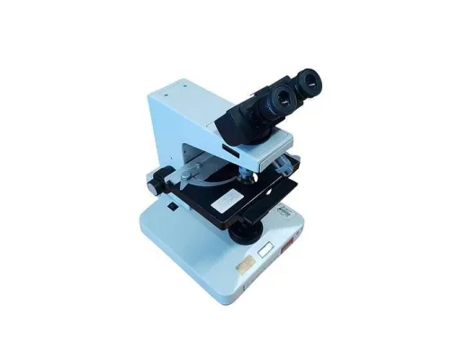
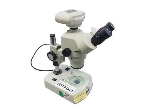
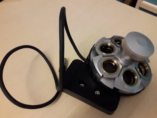
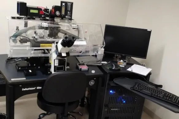
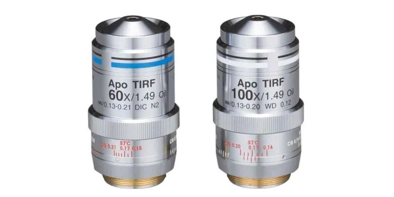
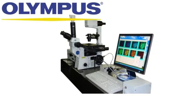
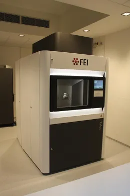
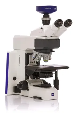
Once a specimen is mounted, the question is how it will be illuminated and magnified. The listings span a wide range of approaches. A conventional compound stand with a set of objectives covers most routine brightfield and fluorescence work. Confocal platforms reject out-of-focus light to build optical sections, which is what lets them reconstruct a volume rather than flatten it. Beyond the optical limit sit the electron instruments: a transmission electron microscope passes a beam through an ultrathin section to resolve internal structure, while atomic force systems trace a surface with a physical probe rather than any beam at all. The choice is set by the scale of the feature, not by preference. A micron-scale cell wants a confocal; a membrane sub-structure wants the transmission instrument; a surface topography at the nanometre level wants the probe. Objectives are listed on their own because they migrate between stands and carry much of the resolving power with them, so a buyer often matches an objective to a problem before matching a body.
Capturing and holding the frame
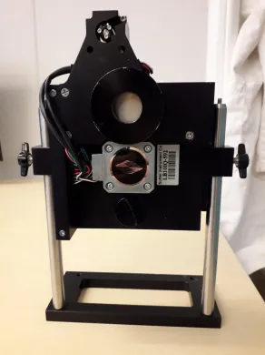
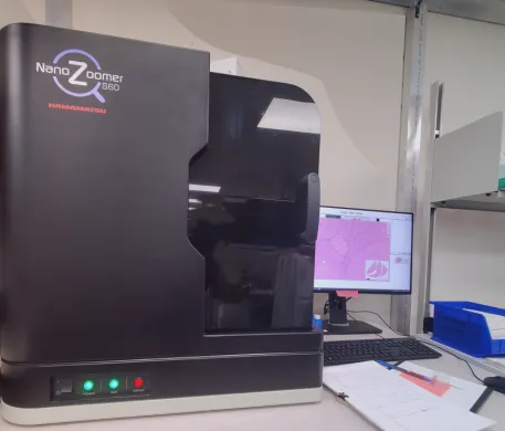
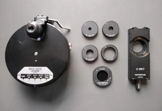
An image that is not recorded stably is lost. Imaging systems in the catalogue cover the detectors, cameras and acquisition hardware that sit at the end of the chain, and microscope accessories cover the stages, holders and optical tweezers that keep a specimen positioned while it is read. This is where mechanical stability stops being an abstraction: a stage that drifts during a long acquisition smears a confocal stack, and a holder that flexes under a probe corrupts an atomic force scan. The categories below are what the marketplace actually carries for this work; among the listings that specify a type, histology and pathology equipment and the imaging systems make up a substantial part of what moves. Where a step in this chain has no category of its own, it is usually folded into one of the broader equipment groups rather than sold apart.
Frequently asked questions
What equipment do you need at minimum to start?
A working chain needs three things: a way to prepare the specimen, an instrument to form the image, and a stable stage and detector to record it. For routine brightfield or fluorescence that can be as little as a compound stand with objectives, a microtome and a slide stainer, plus slides and holders; the preparation step is what sets the quality ceiling, so it cannot be skipped.
Which step is the most expensive to set up?
The image-forming instrument, specifically confocal, electron and atomic force platforms, where the optics, the column and the detector are one integrated purchase rather than a stand with parts added. Preparation equipment like processors, microtomes and stainers sits in the middle, and consumables and single objectives sit at the low end.
What can be bought used and what is better new?
Stands and preparation instruments have mature mechanics, so a used unit often makes sense and is listed below a new one. Electron and confocal platforms are a tighter call because alignment and detector state carry a cost a price tag does not show, so read the individual listing on condition rather than assume.
What has to match between the steps?
Two matches decide the result: section thickness set at the microtome against how much stain can penetrate, and the objective against the detector at the imaging end. A mismatch in either shows up as artefact at the lens or as a frame the detector cannot resolve, regardless of how good the individual parts are.
How much does a complete setup cost?
Across this work prices range from EUR 4 to EUR 244.724, which spans a box of coverslips through to a full electron platform. A routine brightfield or fluorescence chain with preparation equipment falls well below a confocal or beam instrument, so the total depends almost entirely on which image-forming instrument the feature scale requires.
Where does cutting the budget cost you later?
In sample preparation and mechanical stability, not in headline magnification. A block embedded unevenly tears before it sections, an uneven section stains badly, and a stage that drifts smears a long confocal acquisition, so a weak link anywhere in the chain limits everything the lens can deliver.
Which equipment is shared with other work?
Objectives migrate between stands and carry much of the resolving power with them, which is why they are listed on their own and often matched to a problem before a body is chosen. Stages, holders and the general accessories also move between instruments rather than belonging to one body.
What should I check on used equipment for this work?
For stands and preparation instruments, check the mechanics, since that is where wear shows. For confocal and electron platforms, check alignment and detector state, as these carry the hidden cost; the individual listing is where that condition is described, and warranty is arranged in consultation for the specific listing. Parts for these instruments can be sourced via Questpair, which widens what is worth considering second-hand.
About Questpair
Questpair is the European marketplace for lab equipment, where AI-prepared cases and personal specialists guide every purchase. We work with 247 verified suppliers across 38 countries to find buyers the right new, used or refurbished instruments, with response within 1 hour and significant savings vs buying new.
Personal · Fast · Reliable · Affordable