
Histology and pathology equipment
We personally find you the right used, refurbished and new histology and pathology equipment from verified suppliers.
Histology and pathology equipment is the instrumentation used to prepare, section, stain and mount tissue specimens for microscopic examination, covering the full workflow from fixed tissue to a finished slide. The Questpair catalogue holds 309 listings across this class, offered new, used and refurbished by 18 verified suppliers and distributors in 9 countries. 94 percent of the listings are used. These are the instruments a histology or pathology laboratory runs daily: tissue processors, embedding stations, cryostats for frozen sections, automated slide stainers and the consumables that finish a slide. Prices run from EUR 9 to EUR 172.178, a spread that separates a box of coverslips from a fully automated staining platform.
Updated:
Histology and pathology equipment product list
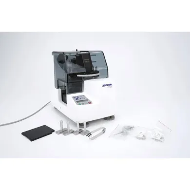
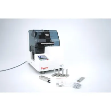


Not the exact instrument you need?
Tell our scientists what you are after. We track it down, including off-market supply.
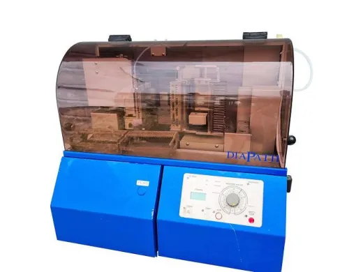
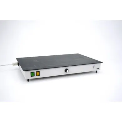
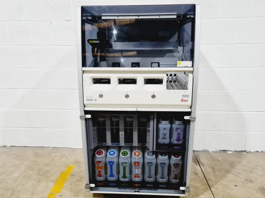
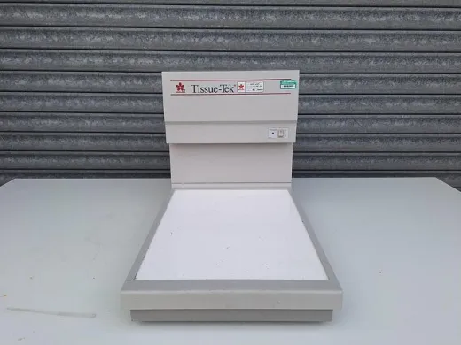
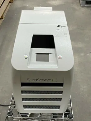
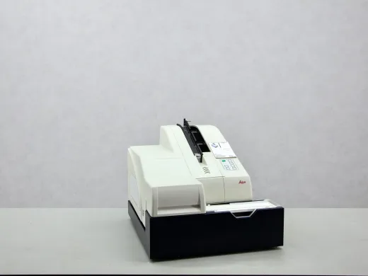
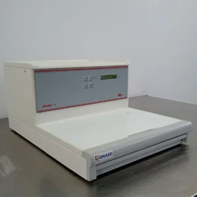
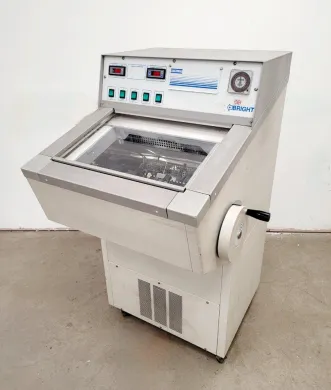
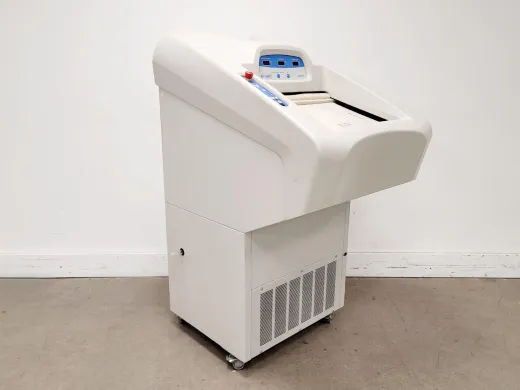
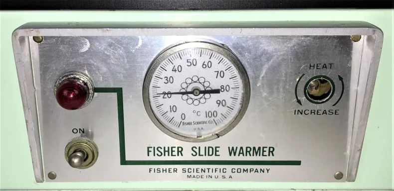
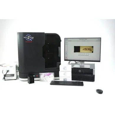
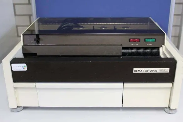
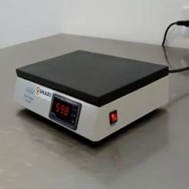
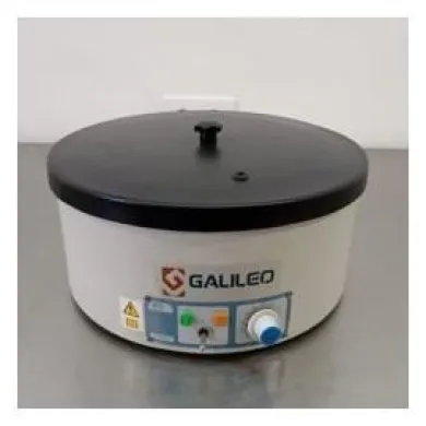
Not the exact instrument you need?
Tell our scientists what you are after. We track it down, including off-market supply.
Showing 1–40 of 216 listings
Show 40 more listingsBuying guide
Throughput settles most histology purchases before any other specification. A laboratory that cuts and stains a dozen blocks a day works to a different standard than one turning over several hundred, and that volume decides whether a manual or a batch-capable instrument fits. A tissue processor rated for 300 cassettes per run suits a hospital pathology service; a 60-cassette benchtop unit suits a research group or a veterinary practice. Match the stated capacity to your real daily block count, not to a peak you reach twice a year, because an oversized processor ties up capital and bench space that a smaller unit would free.
What to verify on a used instrument in this field is specific and worth doing in person. On a cryostat, the chamber cooling and the blade advance are the parts that wear, so confirm the unit reaches and holds its set temperature and that the microtome mechanism feeds cleanly at a known section thickness. On an automated stainer or processor, the reagent handling is what fails: check the pumps, the valves and the fluidics for residue or leaks, and ask whether the unit has run recently rather than sitting dry. A processor left with reagents in its retorts can corrode internally. Recalibration and a fresh set of reagents are a routine cost on any used unit here, so factor them in rather than assuming the instrument arrives ready to run.
The price band is wide because the class spans consumables and capital equipment. Coverslips and slide boxes sit at the bottom, a few euros an item, and a budget stretches a long way at that end. Automated tissue processors and large staining platforms occupy the top, where a single instrument can cost as much as a small vehicle. The average listing sits at EUR 6.110. The brands that dominate the listings are Leica, Abbott Molecular, Leica Biosystems, Thermo Scientific, LAUDA, and their presence matters for a buyer: Leica and Leica Biosystems units are common enough that models can be compared directly and parts are easier to source, which steadies the resale price. A refurbished instrument listed for sale is more affordable than its new equivalent, and for a laboratory outfitting a second bench or replacing a single failed unit, the used market is where most of these purchases actually happen. Prices run from EUR 9 to EUR 172.178.
Types of Histology and pathology equipment
Slide stainers
Slide stainers automate the sequence of reagent baths that bring contrast to a mounted tissue section, moving racks of slides through fixatives, dyes and rinses on a timed program. Automating the sequence removes the variation that hand-dipping introduces, so a batch of hematoxylin and eosin slides comes out consistent from the first rack to the last. This consistency is what makes them central to histology and to the histopathology reporting that depends on reproducible staining. The larger linear and carousel models handle high daily volumes; smaller benchtop units suit a research laboratory running special stains. Among the listings that specify a type, slide stainers are the most numerous subtype here, which reflects how routine automated staining has become in diagnostic pathology.
Embedding stations
Embedding stations are the heated and cooled workstations where processed tissue is set into a paraffin block, with a warm paraffin reservoir, a dispensing nozzle and a cold plate to solidify the mould. The embedding step orients the specimen correctly before it is sectioned, and a station that holds its paraffin at a stable temperature keeps the wax free of bubbles and setting lines. Pathology laboratories buy these in pairs with a tissue processor, since the two define the preparation bench. The distinction from a processor is one of stage: the processor infiltrates the tissue, the embedding station fixes its final orientation in a block ready for the microtome.
Cryostats
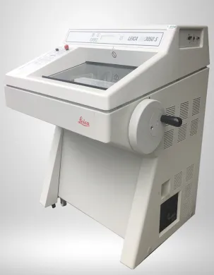
Cryostats are refrigerated microtomes that cut frozen tissue at thicknesses of a few micrometres, enclosing the blade and specimen in a chamber held well below zero. Freezing the tissue rather than embedding it in paraffin is what makes the cryostat fast, and that speed is why it serves intraoperative frozen-section diagnosis, where a surgeon waits for a margin to be read while the patient is still on the table. Cryostats also do immunofluorescence work and molecular diagnostics where paraffin processing would destroy the target antigen. The chamber temperature and the blade feed are the mechanisms that distinguish a good unit; both wear, and both are what a buyer checks first on a used machine.
Tissue processors
Tissue processors carry out the infiltration sequence that replaces the water in a fixed specimen with paraffin, cycling the tissue through dehydrating alcohols, a clearing agent and molten wax over a program lasting several hours. Automating this overnight run is what lets a laboratory present embedded blocks ready for sectioning each morning. These instruments anchor the histology workflow and are sized by cassette capacity, from benchtop units for a research group to enclosed, vacuum-assisted machines for a hospital pathology service. Among the subtypes here they are the fewest in number but the largest in price, since a processor is the instrument a laboratory builds its throughput around.
Coverslips
Coverslips are the thin glass squares laid over a stained section to protect it and to give the microscope a flat, consistent optical surface to read through. They close the slide-preparation workflow, and their thickness is specified to match the objective lenses used for cell imaging and routine light microscopy in histology and microbiology. Although they are a consumable rather than an instrument, they appear in this catalogue because a laboratory sources them alongside its stainers and mounting media. At the bottom of the price range, they are where a limited budget reaches furthest, and they are the subtype a buyer replenishes most often rather than buys once.
Frequently asked questions
What is histology and pathology equipment?
It is the instrumentation used to prepare, section, stain and mount tissue specimens for microscopic examination, covering the workflow from fixed tissue to a finished slide. The class includes tissue processors, embedding stations, cryostats, automated slide stainers and the consumables that close a slide.
What does histology and pathology equipment mean?
Histology is the microscopic study of tissue structure, and pathology is the study of disease through that tissue. The equipment is the set of instruments a laboratory runs to turn a specimen into a readable slide for diagnosis or research.
How does histology and pathology equipment work?
Each instrument handles one stage of a sequence: a tissue processor replaces water in the specimen with paraffin, an embedding station sets it into a block, a microtome or cryostat cuts thin sections, and a stainer runs those sections through reagent baths for contrast. A coverslip then seals the finished slide for the microscope.
What types of histology and pathology equipment are there?
The listings here span histology equipment, automated slide stainers, cryostats, slide processing systems, molecular diagnostics equipment and laboratory automation equipment. Within that, slide stainers, embedding stations, cryostats, tissue processors and coverslips are the subtypes a histology bench uses daily.
What is histology and pathology equipment used for?
It prepares tissue specimens for microscopic diagnosis in hospital pathology and for tissue research. Specific uses include routine hematoxylin and eosin staining, frozen-section diagnosis during surgery, and immunofluorescence or molecular work where paraffin processing would destroy the target.
How do I choose the right instrument?
Start with throughput: match the stated cassette or slide capacity to your real daily block count rather than to an occasional peak. A hospital pathology service needs batch-capable processors and larger stainers, while a research group or veterinary practice is often served by a benchtop unit.
What is the difference between refurbished and used?
A used unit is sold in its existing condition, while a refurbished unit has been reconditioned before resale. On either, confirm what was checked or replaced, since recalibration and fresh reagents are a routine cost on any pre-owned instrument in this field.
How much does histology and pathology equipment cost?
Prices in this catalogue range from EUR 9 to EUR 172.178, with the average listing at EUR 6.110. The spread reflects a class that runs from a box of coverslips to a fully automated staining or processing platform.
Which brands are available in this category?
The brands that appear across the listings include Leica, Leica Biosystems, Thermo Scientific, Abbott Molecular and LAUDA. Leica and Leica Biosystems units are common enough that models can be compared directly and parts are easier to source.
Can I get a warranty on a refurbished unit?
A warranty is often possible and is arranged in consultation for the specific listing. Check the individual listing to see what terms apply before you buy.
Are parts available for these instruments?
Parts can be sourced via Questpair. On established brands such as Leica and Leica Biosystems, the number of comparable units in circulation tends to make sourcing easier.
How long does histology and pathology equipment last?
A capital instrument like a tissue processor or cryostat has a long service life when maintained, but the wear points matter: cryostat chamber cooling and blade feed, and the pumps, valves and fluidics on processors and stainers. Reagent handling left to sit dry or corrode is what shortens a unit's usable life.
Should I buy new or refurbished?
Used and refurbished units are listed at lower prices than new, which is why a laboratory outfitting a second bench or replacing a single failed unit often buys pre-owned. Weigh the two against the individual listing, and note that warranty is arranged in consultation rather than set centrally.
Where can I buy histology and pathology equipment?
The Questpair catalogue holds 309 listings in this class from 18 verified suppliers and distributors across 9 countries. Both new and used stock are listed, with 291 of the listings used.
What are the lead time and shipping like?
Lead time and shipping depend on the individual listing and the supplier's location, which spans 9 countries. Check the specific listing for its availability and dispatch details.
What calibration and maintenance does a used unit need?
Plan on recalibration and a fresh set of reagents as a routine cost rather than assuming the instrument arrives ready to run. On a cryostat, confirm it reaches and holds its set temperature and that the microtome feeds cleanly; on a stainer or processor, check the pumps, valves and fluidics for residue or leaks.
Which types are listed most, and who reaches for which?
Among the listings that specify a type, slide stainers are the most numerous subtype, which reflects how routine automated staining has become in diagnostic pathology. Tissue processors are the fewest in number but the largest in price, since they anchor a laboratory's throughput; embedding stations are usually bought alongside them to define the preparation bench.
Which brands dominate this category, and what does that mean for a buyer?
Leica, Leica Biosystems, Thermo Scientific, Abbott Molecular and LAUDA appear across the listings. Where a brand is common, models can be compared directly and parts are easier to source, which also steadies the resale price of a used unit.
What separates the bottom of the price band from the top?
The bottom is consumables: coverslips and slide boxes at a few euros an item, where a limited budget reaches furthest and the buyer replenishes rather than buys once. The top is capital equipment, automated tissue processors and large staining platforms, where a single instrument can cost as much as a small vehicle, and the gap is one of consumable versus capital rather than quality tiers.