
Imaging systems
We personally find you the right used, refurbished and new imaging systems from verified suppliers.
Imaging systems are laboratory instruments that capture, record and quantify visual or signal-based data from biological and chemical samples, ranging from stained gels and blots to live cells and whole-organism fluorescence. The category spans optical microscopes, gel and blot documentation systems, and molecular imagers that detect chemiluminescent, fluorescent and radioisotopic signals. On the Questpair marketplace the category holds 472 listings from 16 verified suppliers and distributors across 10 countries. 99 percent of the listings are used. New, used and refurbished units all appear, with prices running from EUR 31 to EUR 172.178. A laboratory sourcing imaging equipment can therefore compare a documentation bench against a confocal platform within one catalogue.
Updated:
Imaging systems product list


Not the exact instrument you need?
Tell our scientists what you are after. We track it down, including off-market supply.
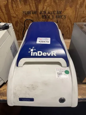
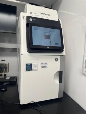
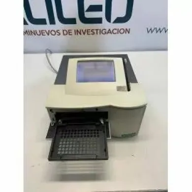
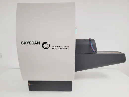
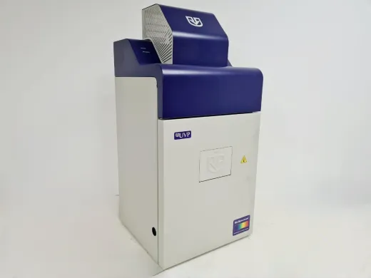
Not the exact instrument you need?
Tell our scientists what you are after. We track it down, including off-market supply.
Showing 1–40 of 271 listings
Show 40 more listingsBuying guide
What settles an imaging purchase before anything else is the signal the instrument has to detect, because that decides the detector, the optics and most of the price. A gel documentation unit reading ethidium bromide under UV is a different machine from a cooled-CCD molecular imager reading faint chemiluminescence, and a confocal microscope resolving subcellular structure is different again. Match the detection mode to the assay first: brightfield and standard fluorescence for routine documentation, high-sensitivity cooled sensors for low-light chemiluminescence and multiplex fluorescence, and point-scanning or spinning-disk optics where spatial resolution in three dimensions is the point. Everything downstream, the light source, the filter sets, the camera bit depth, follows from that single decision.
On a used unit the sensor and the light source are what a buyer has to verify, because they age in ways a datasheet does not show. A cooled camera loses sensitivity as its cooling degrades, and dark-current noise rises; ask for a recent flat-field or a sample acquisition at the longest exposure the lab will use. Mercury and metal-halide lamps have rated hours and dim well before they fail, and LED engines, while longer-lived, still drift. Filter cubes crack and fade, and for a microscope the objective lenses carry most of the value, so their condition and completeness matter more than the stand. Confirm which software licence transfers with the instrument, since acquisition software is frequently node-locked and a transfer can stall a working system for weeks.
Price on this marketplace covers an unusually wide field, from benchtop documentation to research-grade confocal, which is why the range is what it is. Prices run from EUR 31 to EUR 172.178, with an average around EUR 7.032. A documentation system or a simpler fluorescence imager sits near the lower end and is the affordable entry point for a lab on a fixed budget, while a motorised confocal or a high-content platform accounts for the top. The brands occupying these bands, Bio-Rad, Olympus, Invitrogen, Syngene, Thermo Fisher Scientific, each hold a recognisable position: gel and blot documentation and molecular imaging cluster around some of them, microscopy around others. Because the cost of a unit for sale here depends so heavily on which band it falls into, read the price against the detection mode rather than against the headline figure, and let the specification tell you where in the range a given listing honestly belongs.
Types of Imaging systems
Molecular imagers
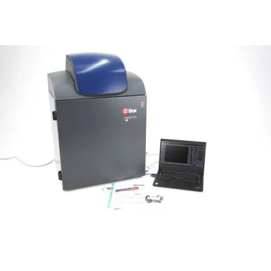
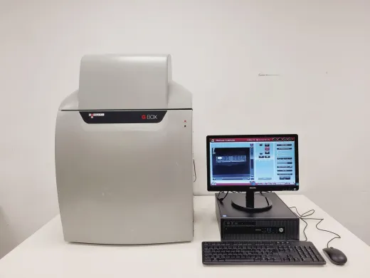
A molecular imager is a cooled-sensor instrument built to capture faint signals from gels, blots and microplates, where the light to be measured is far too weak for an ordinary camera. These systems detect chemiluminescence from Western blots, fluorescence from labelled probes and, in some models, the signal from radioisotopic phosphor screens, acquiring a quantitative image rather than a photograph for the record. The cooled CCD or sCMOS sensor and the long integration times are what separate them from a gel documentation bench: documentation shows that a band is present, a molecular imager measures how much. That quantitative step is why they belong to several research fields at once. They do Western blotting and gel densitometry in protein biochemistry, and the same hardware runs fluorescent and chemiluminescent readouts across molecular biology. In proteomics a molecular imager quantifies protein expression from blots and two-dimensional gels, and in genomics it reads labelled nucleic acids after electrophoresis, so a molecular imager for Western blot quantification and a fluorescence imager for protein expression analysis describe one class of instrument used across these workflows. They are also used for nucleic acid gel analysis and multiplex fluorescent detection where more than one target is read from a single membrane. These systems appear on the Questpair catalogue, and among the current listings 99 percent are used.
Frequently asked questions
What is an imaging system?
An imaging system is a laboratory instrument that captures, records and quantifies visual or signal-based data from biological and chemical samples. The category covers optical microscopes, gel and blot documentation units, and molecular imagers that detect chemiluminescent, fluorescent and radioisotopic signals.
What does imaging system mean in a lab context?
It means any instrument that turns a sample's visual or signal output into a recorded image for documentation or measurement. In practice this ranges from a camera-based gel documentation bench to a cooled-sensor molecular imager that measures faint light rather than just photographing it.
How do imaging systems work?
A light source or signal excites the sample, and a detector such as a CCD, sCMOS or scanning sensor records the emitted or transmitted light. Documentation systems capture a straightforward picture, while molecular imagers use cooled sensors and long integration times to quantify weak chemiluminescent or fluorescent signals.
What types of imaging systems are there?
Listings here include gel documentation systems, fluorescence imaging systems, chemiluminescence imagers, UV transilluminators and gel imaging systems, alongside optical microscopes and molecular imagers. The dividing line is the signal: brightfield and UV documentation, high-sensitivity chemiluminescence and fluorescence, or spatial resolution through confocal optics.
What are imaging systems used for?
They are used for Western blotting, gel densitometry, nucleic acid gel analysis, protein expression quantification and multiplex fluorescent detection. Microscopy platforms add live-cell imaging and three-dimensional resolution of subcellular structure.
How do I choose an imaging system?
Start with the signal the instrument has to detect, because that decides the detector, the optics and most of the price. Match the detection mode to the assay: brightfield and standard fluorescence for routine documentation, cooled high-sensitivity sensors for low-light chemiluminescence and multiplex fluorescence, and point-scanning or spinning-disk optics where three-dimensional resolution is needed.
What is the difference between refurbished and used imaging systems?
A used unit is sold in its current condition, while a refurbished unit has been checked and restored to working order before sale. On either, verify the sensor and the light source, since a cooled camera loses sensitivity as its cooling degrades and lamps dim before they fail.
How much does an imaging system cost?
Prices on this marketplace run from EUR 31 to EUR 172.178, with an average around EUR 7.032. A documentation system or a simpler fluorescence imager sits near the lower end, while a motorised confocal or high-content platform accounts for the top.
Which brands make imaging systems?
Brands listed in this category include Bio-Rad, Olympus, Invitrogen, Syngene and Thermo Fisher Scientific. Gel and blot documentation and molecular imaging cluster around some of these names, and microscopy around others.
Can I get a warranty on a refurbished imaging system?
A warranty is often possible and is arranged in consultation for the specific listing. Terms are set per unit rather than centrally, so check the individual listing.
Are parts available for these imaging systems?
Parts can be sourced via Questpair. For a used microscope the objective lenses carry most of the value, and filter cubes crack and fade over time, so confirm which components are present before you buy.
How long does an imaging system last?
The instrument frame and optics can last many years, but the sensor and light source age fastest. Cooled cameras lose sensitivity as cooling degrades, mercury and metal-halide lamps have rated hours, and LED engines last longer but still drift.
Should I buy new or refurbished?
Used and refurbished imaging systems are listed at lower prices than new units, which is the practical difference on this marketplace. Where you weigh the two, check the sensor and lamp condition on the specific listing and note that warranty is arranged in consultation.
Where can I buy an imaging system?
This category holds 472 listings from 16 verified suppliers and distributors across 10 countries. You can compare a documentation bench against a confocal platform within one catalogue.
What are the lead time and shipping like?
Lead time and shipping depend on the individual listing and the supplier's location across the 10 countries represented. Contact Questpair for the timeline on a specific unit, and note that a node-locked software licence transfer can delay a working system by weeks.
What calibration and maintenance do imaging systems need?
Routine checks include flat-field verification for cooled cameras, lamp hour tracking, and inspection of filter cubes and objectives. On a used unit, ask for a recent flat-field or a sample acquisition at the longest exposure the lab will use to confirm sensor condition.
Which types in this category are listed most, and who reaches for which?
Gel documentation systems, fluorescence imaging systems, chemiluminescence imagers, UV transilluminators and gel imaging systems all appear. Protein biochemists reach for chemiluminescence imagers for Western blots, molecular biologists for fluorescence imagers for labelled probes, and labs running routine gels for documentation systems and transilluminators.
Which brands dominate this category, and what does that mean for a buyer?
Bio-Rad, Olympus, Invitrogen, Syngene and Thermo Fisher Scientific recur across the listings, with documentation and molecular imaging concentrated around some and microscopy around others. For a buyer this means the brand often signals which detection mode a unit is built for, so read the specification alongside the name.
What separates the bottom of the price band from the top here?
The bottom holds benchtop documentation units and simpler fluorescence imagers that read strong signals under UV or standard fluorescence. The top holds cooled-sensor molecular imagers for faint chemiluminescence and motorised confocal or high-content platforms, where the detector, optics and multiplex capability drive the cost. Read the price against the detection mode rather than the headline figure.