
Microscopy equipment
We personally find you the right used, refurbished and new microscopy equipment from verified suppliers.
Microscopy equipment is the class of optical and electron instruments that magnify specimens beyond the resolving power of the unaided eye, together with the objectives, stages, cameras and illumination parts that complete them. The Questpair catalogue holds 7210 listings in this category, drawn from 30 verified suppliers and distributors across 14 countries. New, used and refurbished units are all listed, and 95 percent of them are used, the remainder new. This class of instrument spans simple benchtop light microscopes through to electron and scanning-probe systems, so a buyer meets a wide range of prices and condition grades on a single page. Microscopy sits at the centre of cell imaging, histology and materials analysis in the laboratory.
Updated:
Microscopy equipment product list
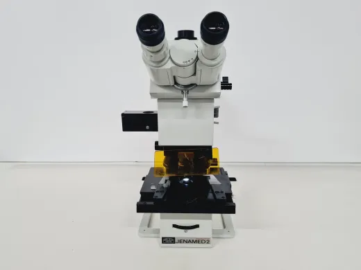
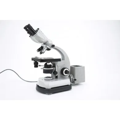
Carl Zeiss Jenamed 2 Microscope with 5 Objectives
- Category
- Objectives
- Brand
- Zeiss
- Suppliers
- 1 verified


Not the exact instrument you need?
Tell our scientists what you are after. We track it down, including off-market supply.
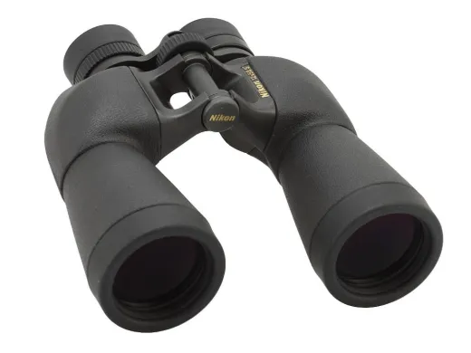
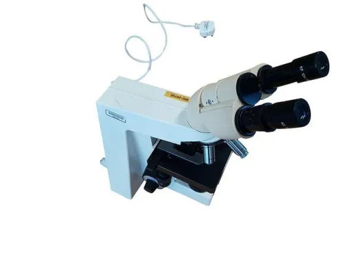
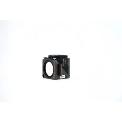
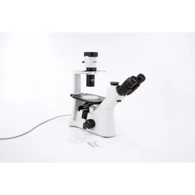
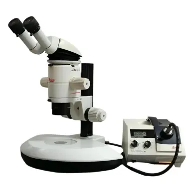
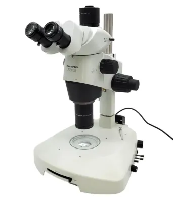
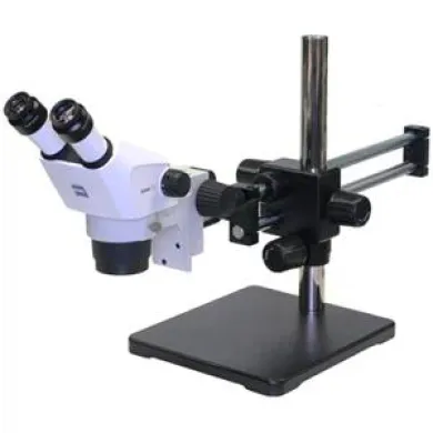
Not the exact instrument you need?
Tell our scientists what you are after. We track it down, including off-market supply.
Showing 1–40 of 6,848 listings
Show 40 more listingsBuying guide
The resolution the work actually needs
What settles a microscopy purchase is the resolving power the sample demands, and everything else follows from it. A routine light microscope resolves to roughly 200 nanometres, which is enough for stained tissue sections, cultured cells and most teaching work, so for histology or microbiology the choice narrows to objective quality, illumination and whether the stand is upright or inverted. Below that limit the question changes entirely: imaging a virus, a grain boundary or a nanostructure moves the buyer to electron or scanning-probe instrumentation, where a transmission electron microscope reaches sub-nanometre detail and a scanning electron microscope delivers depth of field across a rough surface. Confocal systems sit between these worlds, rejecting out-of-focus light to build optical sections of thick fluorescent specimens. Fix the smallest feature that must be seen clearly, and the class of instrument is already decided.
What to verify on a used unit
Because used and refurbished instruments dominate this catalogue, condition is where most of the risk sits. On a light microscope, check that the objectives match the stand and that none show fungus, delamination or scratched front elements, since a replacement objective can cost more than the body. Confirm the illumination source, as older halogen lamps and some arc burners are harder to replace than LED. On electron instruments the stakes are higher: ask about column vacuum history, filament or emitter hours, detector condition and whether the control electronics and software still run on supportable hardware. A used electron microscope that has sat unpowered for years may need vacuum restoration before it images at all. Where a listing is thin on these details, the specialists reachable via Questpair respond within an hour, and parts can be sourced through the same route.
Where the price band sits
Microscopy equipment covers an unusually wide cost range, from EUR 4 for small accessories and single objectives to EUR 236.745 for a complete electron or confocal system, with an average listing around EUR 3.241. That spread reflects the gap between a teaching light microscope and a research-grade analytical instrument far more than any difference in condition. For a buyer working to a fixed budget, the used and refurbished units that make up most of the catalogue are the affordable route into capabilities that are expensive when bought new. Brand matters here too: Olympus, Nikon, Zeiss, Leica, Motic account for a large part of the light-microscope listings, which keeps models easy to compare and parts easier to find. Many of these units are listed for sale at a fraction of their original cost, so the practical question is less what a given instrument costs in isolation than which condition grade and brand fit the work at the price available.
Types of Microscopy equipment
Microscopes
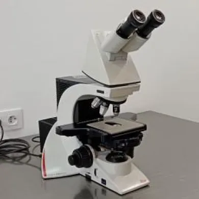
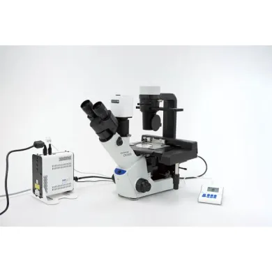
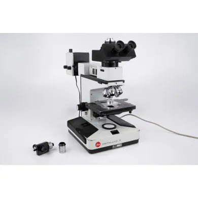
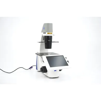
Microscopes are the compound light instruments that form the core of this category, using glass objectives and an eyepiece or camera to magnify specimens illuminated by transmitted or reflected light. They range from basic monocular teaching stands through trinocular research microscopes with phase contrast, darkfield and fluorescence options. The practical division is between upright models, where the objective sits above a stage, and inverted models, where it sits below, which is the configuration cell culture and live-cell work demand because the sample stays in its vessel. This is the equipment behind routine histology, microbiology and cell imaging, and fluorescence-capable units carry much molecular biology and immunoassay work where labelled targets must be located inside a cell. Light microscopes also serve quality control and materials inspection across industry.
Microscope accessories
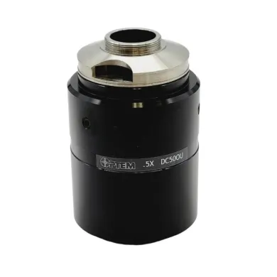
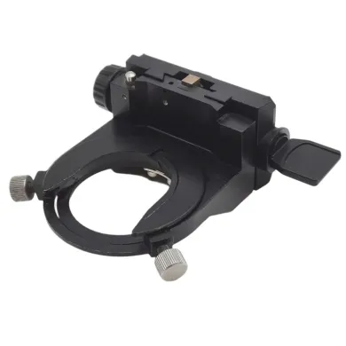
Microscope accessories are the components that extend or complete a microscope rather than image on their own: cameras and adapters, stages and stage inserts, condensers, illuminators, filters, eyepieces and mechanical spares. They are the second-largest group in the catalogue, which reflects how often a working microscope is built up or repaired part by part rather than replaced. For a buyer, this class is where a monochrome research camera, a motorised stage or a replacement fluorescence filter cube is found, and it is the route to keeping an older but optically sound body in service. Accessories also carry the imaging and analysis chain for cell imaging and electrophoresis gel documentation, where the camera and software matter as much as the optics beneath them.
Objectives
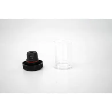
Objectives are the primary lenses that gather light from the specimen and determine a microscope's resolution and image quality, specified by magnification, numerical aperture and the correction class that governs how well they handle colour and field flatness. Plan achromats suffice for routine work, while apochromats and plan-fluorite objectives are chosen where colour fidelity and resolving power matter, as in fluorescence and high-magnification cell imaging. Immersion objectives, used with oil or water, push numerical aperture and therefore resolution higher still. Because an objective can represent a large share of a microscope's optical value, buyers often source them separately to upgrade an existing stand or to replace a damaged lens, matching the thread, tube length and coverslip correction to the body already owned.
Transmission electron microscopes
Transmission electron microscopes pass a focused beam of electrons through an ultra-thin specimen to resolve internal structure down to the sub-nanometre and atomic scale, far beyond any light instrument. They are the tool for imaging the fine architecture of cells and tissues in histology and ultrastructural research, for virology and molecular biology where macromolecular assemblies are studied, and for materials science where crystal lattices and defects are examined directly. A TEM is a substantial installation with a high-voltage column, vacuum system and specialised sample preparation, so listings tend toward research institutions and are priced and serviced accordingly. Buyers evaluate accelerating voltage, resolution specification, detector and camera fit, and the practical state of the vacuum and electronics.
Scanning electron microscopes
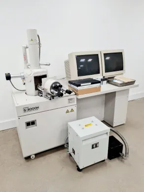
Scanning electron microscopes scan a focused electron beam across a specimen surface and collect the resulting secondary and backscattered electrons to build a high-resolution image with great depth of field. Where a TEM looks through a sample, an SEM looks at its surface, which makes it the instrument of choice for topography, fracture analysis and particle morphology, and, with an energy-dispersive detector, for elemental microanalysis. In the life sciences it supports cell-surface imaging and the examination of tissues and microorganisms, and in materials and failure analysis it is a mainstay. Compact tabletop SEMs have widened the user base beyond dedicated facilities. A buyer weighs resolution, chamber size, detector complement and whether the model works with conventional or variable-pressure modes for uncoated or biological specimens.
Confocal microscopes
Confocal microscopes use a pinhole to reject out-of-focus light, so they build sharp optical sections of thick fluorescent specimens and reconstruct them into three-dimensional images that a conventional fluorescence microscope cannot resolve cleanly. This makes them central to modern cell imaging and molecular biology, where labelled proteins and structures are localised through the depth of a cell or tissue, and to colocalisation and live-cell studies in cell culture. Laser-scanning and spinning-disk variants trade imaging speed against resolution and photobleaching, which is the main distinction a buyer draws. A confocal system bundles lasers, scan optics, detectors and analysis software, so listings are evaluated as a whole, and the laser lines available dictate which fluorophores the system can excite.
Atomic force microscopes
Atomic force microscopes image a surface by tracking the deflection of a sharp probe on a cantilever as it is raster-scanned, producing topographic maps at nanometre and sub-nanometre resolution without any beam or lens. Unlike electron and light instruments, they need no vacuum or staining and work in air or liquid, which lets them image soft biological material, single molecules and living cells under near-native conditions, a reach into molecular biology and nanoscale biophysics that optical methods cannot match. They are equally at home in materials and surface science, mapping roughness, mechanical properties and nanostructures. These systems are the rarest entry in the catalogue, so availability is limited, and a buyer assesses the scan range, available imaging modes and the condition of the scanner and probe holder.
Frequently asked questions
What is microscopy equipment?
It is the class of optical and electron instruments that magnify specimens beyond what the unaided eye can resolve, together with the objectives, stages, cameras and illumination parts that complete them. The range runs from benchtop light microscopes to electron and scanning-probe systems.
What does microscopy equipment mean in practice?
In practice it refers to the whole imaging chain: the microscope body, the lenses that form the image, the light source or electron source, and the camera or eyepiece that captures it. A single working setup is often assembled from several of these parts rather than bought as one unit.
How does microscopy equipment work?
Light microscopes pass or reflect visible light through glass objectives to magnify a specimen, while electron instruments use a focused electron beam that either passes through a thin sample or scans its surface. Scanning-probe microscopes such as atomic force models track a physical probe over a surface instead of using a beam or lens.
What types of microscopy equipment are there?
The main types are optical microscopes, microscope accessories, objectives, transmission electron microscopes, scanning electron microscopes, confocal microscopes and atomic force microscopes. Within optical instruments you will find binocular, trinocular, research and laboratory configurations in upright and inverted forms.
What is microscopy equipment used for?
It supports cell imaging, histology, microbiology and molecular biology in the life sciences, and materials analysis, failure analysis and quality control in industry. The specific instrument depends on the smallest feature that must be resolved.
How do I choose the right microscopy equipment?
Start from the resolution the sample demands: a light microscope resolves to roughly 200 nanometres, which covers stained tissue and cultured cells, while features below that require electron or scanning-probe instruments. Then match objective quality, illumination type and whether an upright or inverted stand suits the work.
What is the difference between refurbished and used microscopy equipment?
Used units are sold in their existing condition, while refurbished units have been checked and brought back toward working order before listing. For both, verify the condition details against the individual listing rather than assuming a grade.
How much does microscopy equipment cost?
Listings range from EUR 4 for small accessories and single objectives to EUR 236.745 for a complete electron or confocal system, with an average around EUR 3.241. The spread reflects the gap between a teaching light microscope and a research-grade analytical instrument more than condition.
Which brands are common in this category?
Olympus, Nikon, Zeiss, Leica and Motic account for a large part of the light-microscope listings. Because these names appear often, models are easier to compare side by side and matching parts are easier to find.
Is there a warranty on refurbished microscopy equipment?
A warranty is often possible and is arranged in consultation for the specific listing. Check the listing itself and raise the question before purchase, since terms vary by unit.
Can I still get parts for microscopy equipment?
Parts such as objectives, stages, condensers, illuminators, filters, eyepieces and camera adapters can be sourced via Questpair. Matching a part correctly means checking thread, tube length and coverslip correction against the body you already own.
How long does microscopy equipment last?
Optically sound microscope bodies can stay in service for many years because they are repaired and built up part by part rather than replaced, which is why accessories form a large group in this catalogue. Electron instruments depend on the state of the vacuum, the filament or emitter hours, and whether the control electronics still run on supportable hardware.
Should I buy new or refurbished microscopy equipment?
Used and refurbished units are listed at lower prices than new, which is the affordable route into capabilities that are expensive bought new. Where you need to weigh the two, compare the condition details on the individual listing and arrange any warranty in consultation.
Where can I buy microscopy equipment?
The Questpair catalogue holds 7210 listings in this category from 30 verified suppliers and distributors across 14 countries. New, used and refurbished units are all listed on the same page.
What are the lead time and shipping like?
Lead time and shipping depend on the individual listing and the supplier handling it, so they are set per unit rather than centrally. Electron instruments in particular are substantial installations, which affects how they are moved and set up.
How is microscopy equipment calibrated and maintained?
Maintenance on a light microscope centres on clean optics, a working illumination source and objectives free of fungus, delamination or scratches. Electron instruments need attention to column vacuum, detector condition and emitter life; Questpair does not carry out servicing, but parts for upkeep can be sourced through it.
Which types in this category are listed most, and who reaches for which?
Optical microscopes form the core of the category, with binocular, trinocular, research and laboratory configurations, and microscope accessories are the second-largest group because working bodies are built up part by part. Histology and microbiology labs reach for light microscopes, cell and molecular biology work reaches for fluorescence and confocal systems, and ultrastructural or materials research reaches for electron and atomic force instruments.
Which brands dominate this category and what does that mean for a buyer?
Olympus, Nikon, Zeiss, Leica and Motic make up a large share of the light-microscope listings. For a buyer this means easier model-to-model comparison and better odds of finding compatible objectives, stages and spares to keep an existing stand in service.
What separates the cheapest microscopy equipment from the most expensive?
At the low end sit small accessories, single objectives and teaching light microscopes resolving to around 200 nanometres, priced from EUR 4. At the high end sit complete electron and confocal systems reaching sub-nanometre detail with lasers, detectors and analysis software, up to EUR 236.745; the difference is the resolution and the imaging chain the work requires, not condition alone.